Carimas is a general medical imaging processing platform developed in Turku PET Centre in Finland.
Originally, Carimas was designed for visualization, segmentation and modelling of PET data only. However, the latest versions support processing of imaging data from most medical imaging modalities, such as CT and MRI.
Using Carimas, you can easily visualize your imaging data in many different ways, such as in 2D or 3D, or fuse images from different modalities (PET/CT, PET/MRI).
Furthermore, Carimas provides a lot of advanced functions for researchers. For example, using ROI/VOI tools, the user can draw a region/volume of interest in manual, semi-automatic or automatic manner; using the modelling tools, the user can perform advanced analysis for his/her research data; using Heart tools, cardiac researchers can easily analyze their PET studies.
Features
Input
-
Multiple image data format support: DICOM, ECAT, Analyse, Interfile, Nifti, Interfile, MicroPET and general bitmap formats (JPG, TIFF, PNG and BMP).
-
PACS support: available to connect to hospital PACS system.
Visualization
-
View images from transaxial, coronal, sagittal or any free direction.
-
3D view with color rendering or MIP.
-
Move and rotate images freely in 3D space.
-
Visualize image histograms or cut profiler lines.
Static image tools
-
Calculate VOI statistics: mean, maximum, minimum, standard deviation, volume, etc…
-
SUV and percentage units.
Dynamic image tools
-
Easy and fast visualization of dynamic data.
-
Calculate sum or difference images, or view individual frames.
-
Analyze time activity curves from VOIs or individual voxels.
Segmentation tools
-
2D ROI sets or 3D VOIs.
-
Histogram tool for selecting voxels at value range.
-
Start region definition from some predefined 3D shape or draw your own.
-
Create VOIs using masking and countour tools.
-
Some 3D region growing segmentation tools exist also as a separate plugin, like “syringe” and threshod tools.
-
Scale, move rotate, smooth, combine the VOIs with easy visual tools.
-
Save and reuse the VOIs in multiple studies, regardless of image type or resolution.
Image fusion
-
File format independent image positioning
-
Coregistrate PET/MR/CT automatically
Modeling
-
Use data from images or data files
-
Specify model parameters values and limits
-
Rescale data, define time ranges and exclude time points
-
Calculate parametric images from any model and parameter (separate plugin)
Existing models (trial has only first two)
-
Linear regression
-
Exponential fitting
-
Patlak
-
Logan
-
FUR index
-
Tracer specific models for water (with special license), ammonia, rubidium, acetate and flurpiridaz
-
Generic compartment models
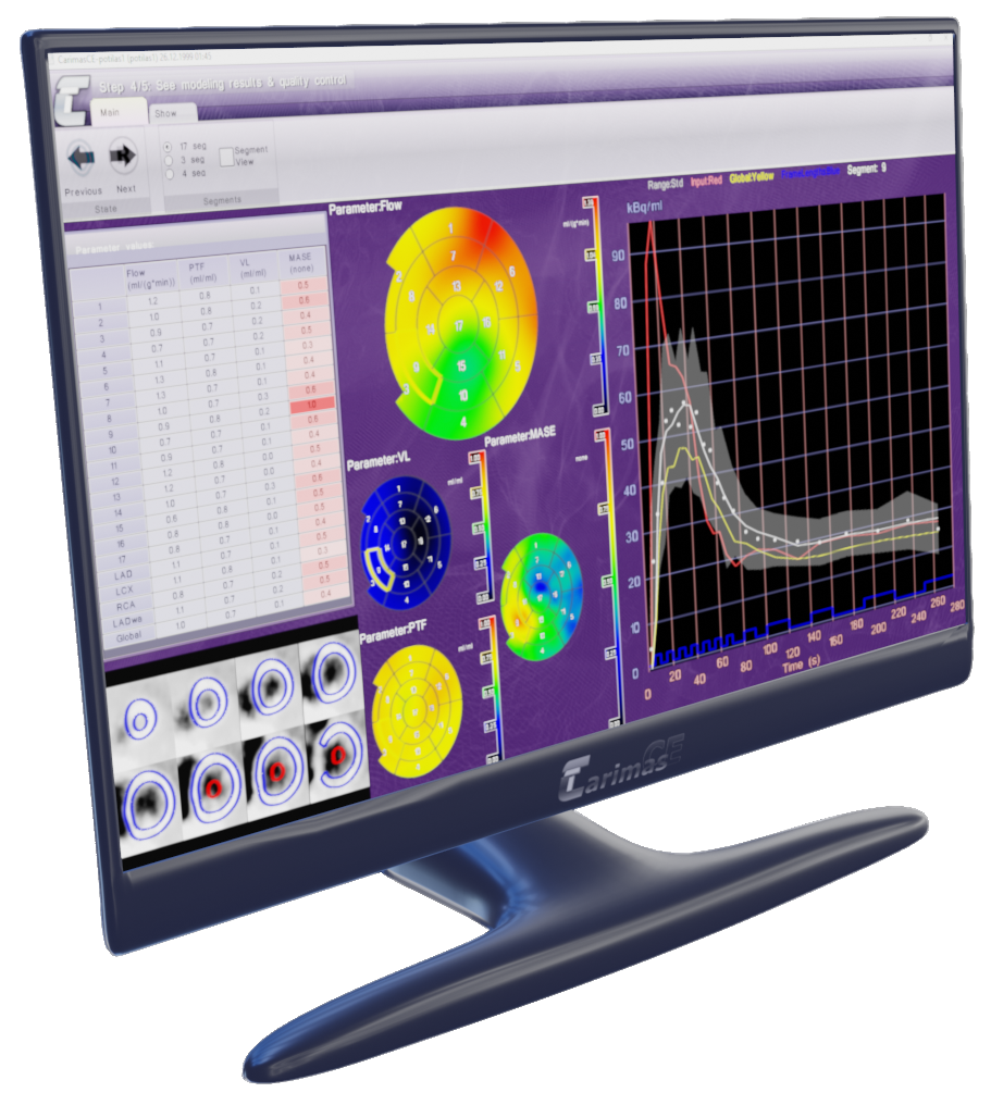
Tools for heart analysis
-
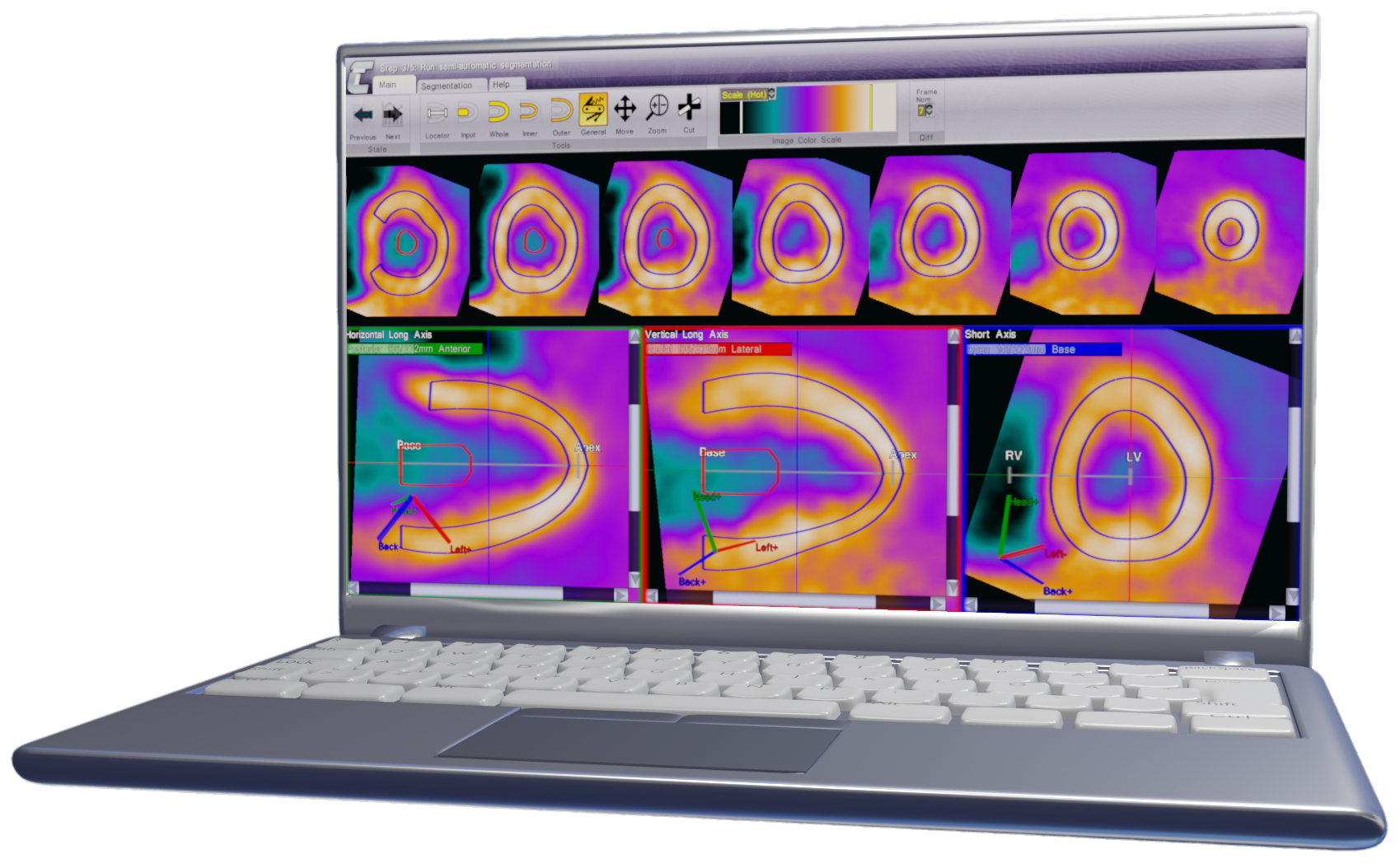
Semi-automatic heart segmentation with manual modification tools
-
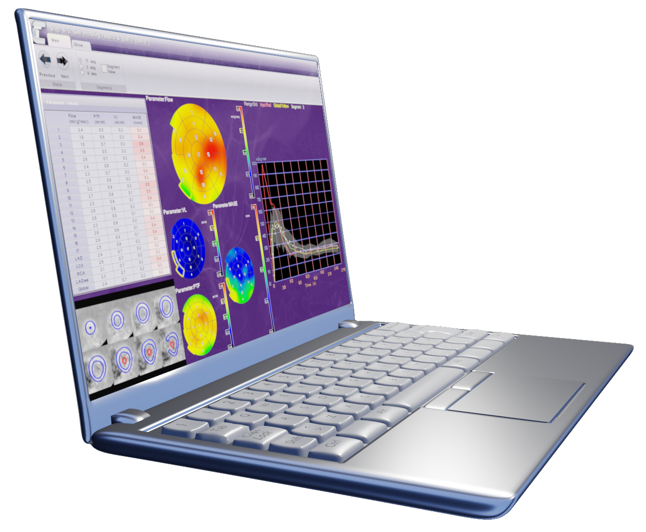
Analyze polarmaps in 3, 4 or 17 segment modes, pixel by pixel or draw your own ROI to polarmap
-
Use any models from the Carimas modelling library to create parametric polarmaps
-
Compare results side by side or save to data files
Lots of other features as plug-ins
-
Add to program easily from Carimas menu by just checking them from list
-
List of currently existing plug-ins can be found here: Go to external plugins archive
Download and Pricing
It is possible to try free trial version of the program for a limited time by downloading the package below and requesting free trial license from the help menu of the program. Please include “trial request” in the subject of the e-mail. We provide one free trial license for each user.
Download Carimas Research trial version.
Pricing
An offer for the full version of the program can be requested through email: sales@carimas.fi
System requirements:
.NET 4.0+ for (2.10) or 2.0+ for older
Older versions
The older program versions than 2.10 can be used by installing version 2.10, running the program and doing following things:
- Select from the menu: Help->Check Updates
- If the program says that there is no updates, click: “Advanced View”
- Check the “show all versions (also old)” -checkbox
- Downgrade the Core version to some of the older versions by clicking the red version number -button. The package versions can be seen in the description window on right when the Core is selected
- Close the module window and restart Carimas
- When program asks to update newest version, select: “Don’t install”
Manuals
Old version 2.4:
General users guide for 2.4 (pdf)
Heart analysis guide for 2.4 (pdf)
External plug-in archive
There exists several additional plug-ins that can be downloaded and added to Carimas. Since version 2.9 all the plug-ins are handled trough built in plug-in manager which can be found from help menu of Carimas. List of currently existing plug-ins can be found here: Go to external plugins archive
NOTE: These do not belong to official Carimas and we give no guarantee of results calculated with these.
If you use the software for research, please cite this article:
Rainio, O., Han, C., Teuho, J. et al. Carimas: An Extensive Medical Imaging Data Processing Tool for Research. J Digit Imaging (2023). https://doi.org/10.1007/s10278-023-00812-1
Roadmap
CORE 2.10.5.0
- Feature: VOI exclusion, focusing and organizing
- Feature: White and dark color themes
- Feature: Custom slice count to plane windows
- Feature: Image focusing
- Feature: Orthonormalizing moves to closest straight orientation
- Feature: Half sphere added
- Feature: Colored background reference images can be added, cleared and edited with GUI
- Feature: The image header dialog allows copying content to clipboard
- Feature: Sum and Sum/volume -columns added to static image display
- Feature: DICOM images transfer syntax can be seen from header dialog
- Feature: Autoradiography HUE -color scale was added
- BugFix: The flipped mesh normals of marching cubes
- BugFix: The invisible Redo -button issue was fixed
- BugFix: The saving forms no longer list wrong formats as options
- BugFix: Non functioning command menu was fixed
- BugFix: The crashing license ordering form fixed
- BugFix: Main – Background toggle shortcut fixed
- BugFix: Non functioning series description fixed from image header dialogs header
TPC library 0.15.6
- Feature: The transfer syntax list of DICOM CStore client can be configured and in Carimas it no longer accepts the unsupported transfer syntaxes
- Feature: The DICOM tag numbers are now shown along with the tag names and values
- Feature: Added UIDs to SRTM and TRTM models, because they were causing complainments in Carimas (having same UID)
- Feature: Added clear messages in cases when non supported data is opened: Packed data, enhanced formats and RGB data
- BugFix: BAS -format scalings are changed to use G values 256 and 65536 instead of 255 and 65535
- BugFix: 8-Bit Bas2500 files can be opened now correctly
Core updates
- Scripting, macro and patch processing support
- More detailed functionality trough manual commands
- Image time shifting
- Uniform image scaling
- Hot colorscale
- RGB background images
- Mask region loading and saving
- Data can be imported to program with drag and drop
- Better logging system
Core updates
- Informs about new available updates and updates if user accepts
- Supports adding analysis flow plug-ins
- Better display for single slice images
- Multiple VOIs can be edited same time as a group
- Already drawn VOIs can be set visible when new regions are drawn
- From menu, it’s possible to query list of plug-ins from Carimas server and install them with a single click
- Support for dynamic masks
Core updates
- Hint paths (projects know now relative paths to images)
- 64-bit indexing (64 bit OS can load image of unlimited size if there is enough memory)
- More plug-in interfaces
- PACS moved to TPC library and updated faster + nearline wait support
- Memory leak bug fix
- 3D masking bug fix and optimization for memory handling
- Tooltip helps added to model tree panel
- Showing and hiding window components
- Possibility to show image information texts on windows (and customize the shown info)
- Screen shots with custom resolution
- Mask image saving with more than one regions
- Converting drawn 2D ROI sets to closed 3D shape. Gaps are interpolated (No need to draw to every plane)
- Cubic interpolation option added for visualization
- Better color bar (value histogram and context menu added)
- Better histogram tool
- Mask transparency slider
- Weighted mean display in addition to sum and diff
- Possibility to display 3D window in free dragball mode (no fixed up vector for camera)
- 2D ROI editor allows now deleting and adding points after ROI is created
- Min/Max curve calculation
Core updates
- Licensing
- Registration plugin interface
- Image orientation bug fix in PACS and Secondary library loading
- Linux tooltip fix and optimization for 64 bit windows
TPC library updates
- Deadlock fix in DicomHeader
- Add bfsrtm
- Use exact match in TextHeader fields
- Add lambda values to Isotope
- Add count information to ImageHeader interface
- Fix WeightedSumOfSquaresMetric
- Always read matching sif file from the image directory, if found
- Add Lambda/HalfLife conversions to Isotope
- Fix DFT.Convolution
- Add constants to Mathematics
- Move LinearEquations to Mathematics
- Add Mathematics namespace
Automatic PET–>MRI coregistration was added
Core updates
- XOR masking for VOIs
- %ID/mL -output unit added
- Now both main image and background can be moved and rotated
- It’s now possible to extract curve from every pixel inside of VOI to file
- Possibility to scale selected VOIs by given percentage
- Possibility to rotate selected VOIs by given angle
- Contour ROI works now with images with one plane
TPC library updates
- Remove info messages to log from Logan and Patlak
- Fix CommonBitmap and Bas file saving as Dicom
- Fix bitmap recognition
- Fix bitmap saving as Dicom
- Fix automatic reading of Sif in Analyze files
- Add IScaling interface; other than linear scaling now possible
- Add Fuji BAS2500 file format
- Fix interfile frame times
- Add orientation info to MicroPET (using subject_orientation)
- Fix CreateDicom when input data is in format not supported by Dicom
- Update fit process optimization logic to ensure fits are not dependent on previous
results - Change Kmono parameter limits
- Fix null pointer crash when opening an empty file as dft
Core updates
- Plot time range selecting accepts now negative values
- Color bar memory consumption bug fixed
- 2D ROI in rotated image crash bug fixed
- VOI copy paste bug fixes
- Plugin folder selecting
TPC library updates
- Use image center as MicroPET origin
- Fix bitmap type recognition
- Fix compartment model input function handling, if input and measured data are not
in the same unit / timescale - Add ErrorFunction to Statistics
- Add common bitmap image read support
- Fix overflow in header ascii strings
- Fix dynamic Ecat63 reading and add frame times
- Fix over/underflow when converting from VAX-floats
- Add special case for dynamic CT images in Dicom
- Add new statistical tools
- Fix saving to PACS / Dicom uid generation / patient birthday dicom encoding
- Ensure Dicom UIDs are not too long
New module was added: HeartROI.dll – User can now draw custom ROI to any heart polarmap
Core updates
- Project files do not require folder any more
- Mesh stabilize tool added
- Multiple simultaneous VOI moving in mm
- Mask VOIs
Data Tree updates
- If some type cannot be created during loading, error is shown instead of breaking
whole process
TPC library updates
- Add R values to linear plots
- Fix NaN checking
- Fix: Image writing/rescaling
- Change variable & function names for consistency
- Fix handling of powell deltas with infinite/NaN parameter bounds
- Check QFac in Nifti: always return 1 or -1
- Fix Patlak/Logan units
- Add DecayCorrection to ImageHeader
- Add new interfile headers for HRRT
- Fix Nifti orientation
- Fix dicom study/series/instance UID generation
- Interfile now uses start of the data file name as study description if no other
available
New module was added: Time Correction Plugin (1.0.0.8459)
Core updates
- Better memory management. Larger images can now be loaded
- Free image rotation support
- Neurological/Radiological viewing conventions
- Images are now loaded to standard orientation instead of data save order
- Direction arrows added to graphical screens
- Modeling parameters can be fixed to one value
- SUV calculation
- Points can be excluded from curves before modelling
- Curves can be saved with Start+End times if available
Data Tree updates
- Project file conversion system
New module was added: PolarMapCalculator.dll – Any of existing polarmaps can now be compared (diff and ratio polarmaps)
Core updates
- Fixed masking bug on left border of image
- VOI Flipping added to VOI panel
- Brain mirror
- Improvements to 2D active contour
- The currently selected pixel value in now better updated status bar (also mouse
roll, etc) - Display shift between main and background images with Ctrl + Shift + LMouse drag
- Many VOI masks can now be visualized same time by selecting many VOIs from list
PACS communication library updates
- Faster and sorted tree view
- Log level added to log events
- Empty Patient ID’s search bug fix
Data Tree updates
- Multi items are now listed better when tree is visualized
- Logging added
- Bug that caused possible freeze when deleting is fixed
Core updates
- Automatic 2D/3D VOI finding
- 3D Mesh filters added to Tools menu: Enlarge, Shrink, Smooth and Simplify
- Images can now be written to disk in DICOM format
- New image location is asked, if the image is not found during project loading
- Time correction plugin added (image filter)
- Several image loading bugs fixed
- Better image header dialog
- Curves are now automatically converted to image units when loading from a disk (if
image units are Bq or kBq) - Project file can now load images with “multiple extensions”
- Toggle between full and vertex mode with Z key
- Vertex mode radius can be adjusted now with Left Mouse Button + Roll
- Fixed 3D VOI masking bug on plane 0
- SIF file support added: User can now read new image times from image sub menu
- Fixed is the bug when removed images were still consuming memory
- 2D ROI sets can now be combined
- A Tooltip bug of VOI context menu is fixed
- Local settings file is now loaded from a program folder if it is not found from
a personal folder
Core updates
- Max curve calculation added
- Dynamic CT images can now be loaded
New module was added: ParametricImageFilterPlugin.dll – Parametric image can be calculated using any existing model
Core updates
- Bug fixes to static image loading with secondary DICOM library
Core updates
- PACS support
- Secondary DICOM library
- Better memory handling for DICOM images loaded with secondary library
- Better logging system
New module was added: PACS communication library – Images can now be loaded from PACS
First release